Details on tiny creatures and organisms that can’t be seen by the naked eye can be absolutely gorgeous — or completely terrifying.
Nikon recently released winners and notable entries for its Small World Photomicrography Competition, and it includes incredibly detailed photos of things like an ant’s face, a gecko’s hand, breast tissue and algae. See some of the images below.
Recommended Videos
The image of the Madagascar giant day gecko’s hand was named the winner for this year, according to Nikon. Grigorii Timin made the image by using high-resolution microscopy and stitching together more than 300 tiles to create the final image. Timin is supervised by Dr. Michel Milinkovitch at the University of Geneva.
“Preparing the sample was an added challenge. Timin performed whole-mount fluorescent staining and tissue clearing to capture the entire embryonic hand with a confocal microscope,” the release states.
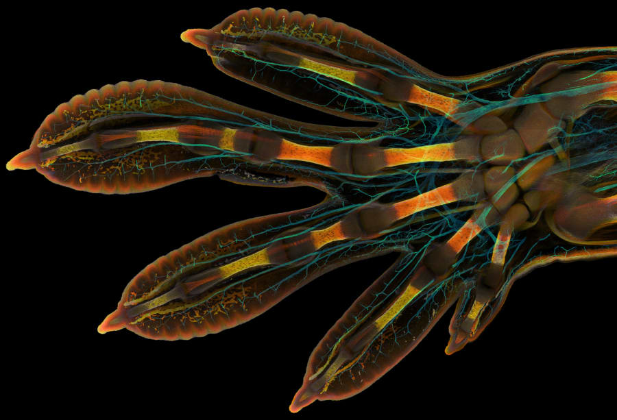
Timin said the gecko’s hand is about 3 mm in length. The photo, which took more than two days to acquire, shows the gecko’s nerves, bones, tendons, ligaments, skin and blood cells.
“This particular image is beautiful and informative, as an overview and also when you magnify it in a certain region, shedding light on how the structures are organized on a cellular level,” Timin said in the release.
The other winners in the top five include:
- Caleb Dawson with an image of breast tissue showing contractile myoepithelial cells wrapped around milk-producing alveoli.
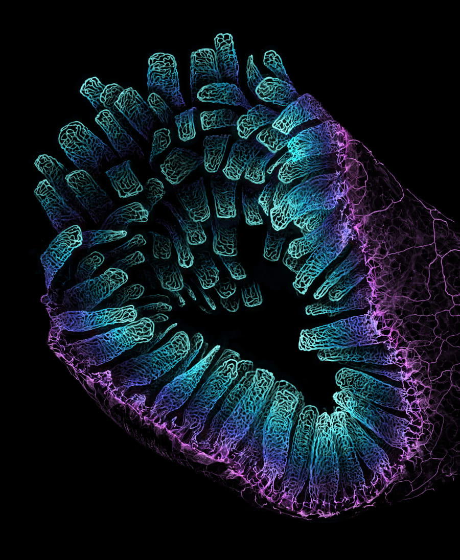
- Satu Paavonsalo and Dr. Sinem Karaman with an image of blood vessel networks from the intestine of an adult moose.
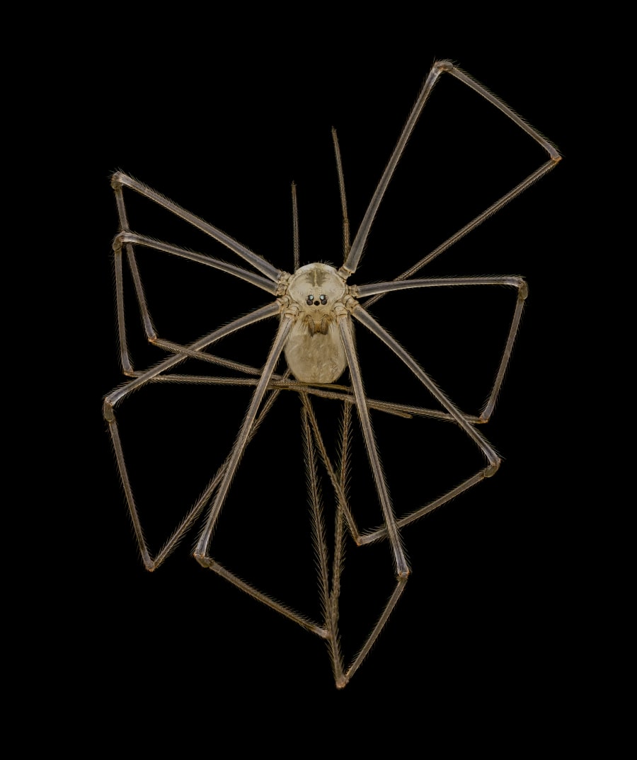
- Dr. Andrew Posselt with an image of a daddy-long-legs spider.
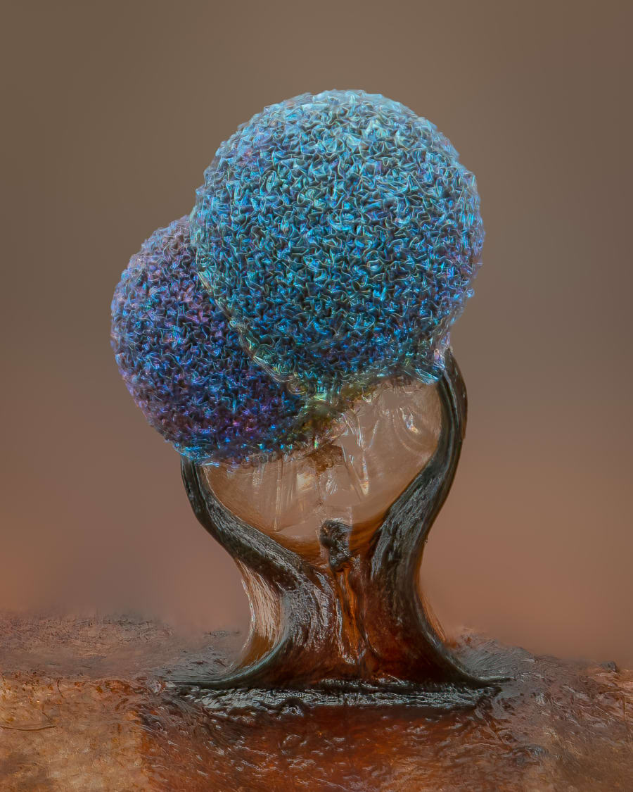
- Alison Pollack with slime mold.
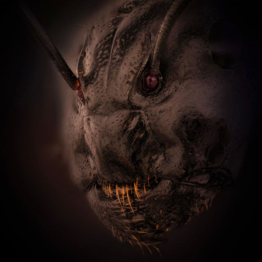
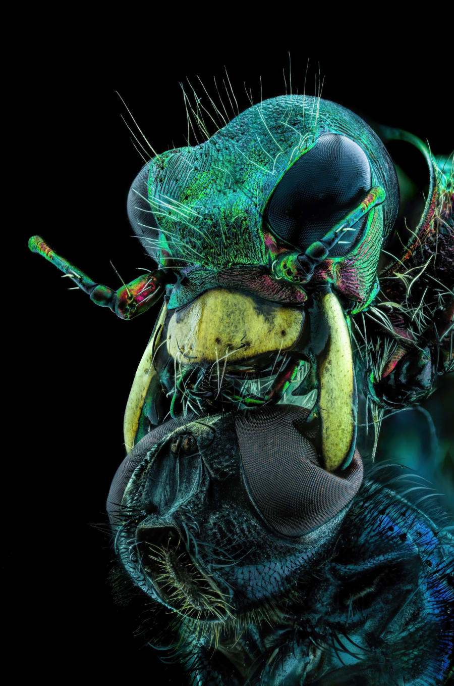
While Timin’s image is stunning, others like the ant’s face are horrifying.
Dr. Eugenijus Kavaliauskas in Lithuania created an image of the ant’s face that captures its reddish eyes, antennas and hair. The image didn’t make the top 20 or honorable mentions though; it was listed as an image of distinction.
This is the 48th annual competition from Nikon. Nikon Small World recognized the photos of 89 artists and scientists out of thousands of submissions.

Here are other images that received recognition.